Dermatologie » Entzündliche Dermatosen » Akne und Rosacea

»

EADV: AI-driven profiling of rosacea severity and markers in clinic and research

Digitale Hände halten leuchtenden AI-Mikrochip – Symbol für Künstliche Intelligenz und Digitalisierung

Quelle: © AiInfo - stock.adobe.com

EADV: AI-driven profiling of rosacea severity and markers in clinic and research

Fachartikel

Dermatologie

Akne und Rosacea

mgo medizin Redaktion

Verlag

3 MIN

Erschienen in: DermaForum

Objectively assessing rosacea severity is difficult. A study demonstrates an AI approach for standardized, quantitative profiling that goes beyond the mere detection of redness.

Rosacea is a chronic inflammatory skin disease in which objective and reproducible assessment of disease severity remains challenging in both clinical practice and research. In this study we presented an AI-driven imaging approach for standardized, quantitative profiling of rosacea severity beyond visible redness alone.

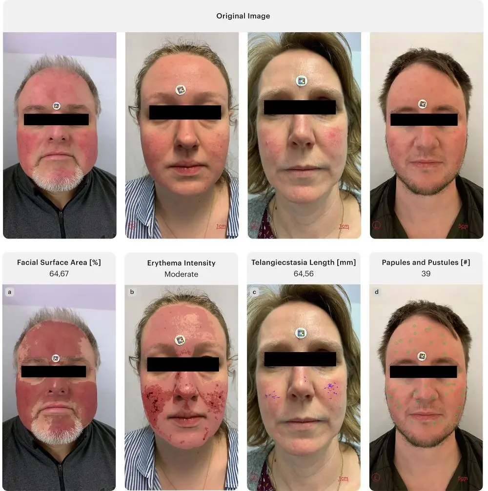
Using the clinically validated Scarletred® Vision Software Platform in combination with a mobile application and standardized image acquisition via a skin patch, facial images were collected longitudinally from 21 patients (355 images). The analysis is powered by Scarletred® ARORA AI-Agent, a multimodal artificial intelligence engine that coordinates image preprocessing, automated feature extraction, and longitudinal pattern analysis. The AI-Agent automatically quantified multiple rosacea-related morphological markers, including affected facial surface area, erythema intensity, telangiectasia length, and number of papules and pustules.

“With this work, our aim was to move beyond Subjective visual grading and provide dermatologists with an objective, standardized tool for assessing rosacea severity. Our AI-derived morphological biomarkers may support future digital endpoints in clinical trials and enable more personalized treatment strategies.”

Chiara Agostini, MSc., Data Scientist

The AI-based analysis accurately delineated erythematous regions, enabling objective measurement of erythema extension and intensity, while simultaneously detecting and quantifying telangiectatic structures, inflammatory lesions, and estimating the presence and severity of rhinophyma. Longitudinal analysis allowed visualization of erythema intensity dynamics and changes in morphological markers over time, supporting precise monitoring of disease progression and treatment response.

BU: Composite figure showing (a) the original image, (b) the corresponding SEV signal map. (c) the graphical representation of the erythema intensity trend; © Supplementary graphics and patient images, with source attribution (SCARLETRED Holding GmbH, Vienna, Austria, and Department of Dermatology, Universitätsklinikum Tuebingen, Tuebingen, Baden-Wuerttemberg, Germany).
Abb.1: Composite figure showing (a) the original image, (b) the corresponding SEV signal map. (c) the graphical representation of the erythema intensity trend; © Supplementary graphics and patient images, with source attribution (SCARLETRED Holding GmbH, Vienna, Austria, and Department of Dermatology, Universitätsklinikum Tuebingen, Tuebingen, Baden-Wuerttemberg, Germany).

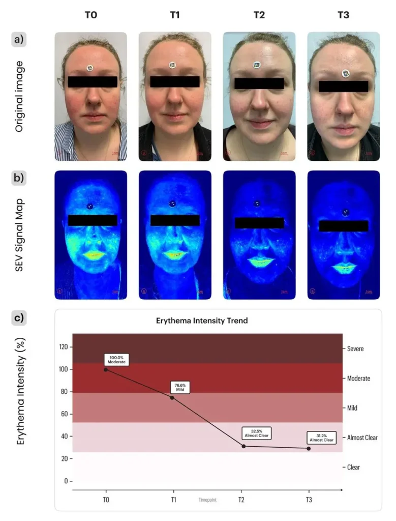
Our results demonstrate the potential of AI-enhanced facial imaging as a standardized, automated, and reproducible framework for rosacea assessment, paving the way toward more personalized and data-driven dermatological care. Our AI-supported framework enables objective, longitudinal rosacea assessment suitable for both routine clinical practice and decentralized monitoring. Unlike conventional ordinal grading systems, it provides continuous, quantitative metrics across multiple morphological dimensions, supporting reproducible digital clinimetry while minimizing inter- and intra-observer variability.

Representative images corresponding to the evaluation of specific clinical parameters. Each image highlights one feature: affected area (a), erythema intensity (b), telangiectasia c), and papules/pustules (d); © Supplementary graphics and patient images, with source attribution (SCARLETRED Holding GmbH, Vienna, Austria, and Department of Dermatology, Universitätsklinikum Tuebingen, Tuebingen, Baden-Wuerttemberg, Germany).

Authors: C. Agostini, M. Molnarova, M. Schaller, and H. Schnidar

Correspondence address:

Chiara Agostini, MSc., Data Scientist

Chiara Agostini, MSc
SCARLETRED Holding GmbH
Wien, Austria

Website: https://www.scarletred.com/

E-Mail: chiara.agostini@scarletred.com


Relevant links to current projects and research related to this work

Schaller, M., Riel, S., Bashur, R., Kurup, N., Schnidar, H., & Fehrenbacher, B. (2022). Ivermectin treatment in rosacea: How novel smartphone technology can support monitoring rosacea-associated signs and symptoms. Dermatologic Therapy, 35(11), Artikel e15869. https://doi.org/10.1111/dth.15869

Ranjan, R., Partl, R., Erhart, R., Kurup, N., & Schnidar, H. (2021). The mathematics of erythema: Development of machine learning models for artificial intelligence assisted measurement and severity scoring of radiation induced dermatitis. Computers in Biology and Medicine, 139, Artikel 104952. https://doi.org/10.1016/j.compbiomed.2021.104952

Partl, R., Jonko, B., Schnidar, S., Schöllhammer, M., Bauer, M., Singh, S., Simeckova, J., Wiesner, K., Neubauer, A., &

Schnidar, H. (2017). 128 SHADES OF RED: Objective remote assessment of radiation dermatitis by augmented digital skin imaging. Studies in Health Technology and Informatics, 236, 363–374: https://pubmed.ncbi.nlm.nih.gov/28508819/

Weitere Beiträge zu diesem Thema

Nahaufnahme von Psoriasis Plaques am Ellbogen einer Person als Symbol fuer die Rolle von PA2G4 bei Psoriasis

Die Rolle von PA2G4 bei Psoriasis

Fachartikel

Neues Target bei Psoriasis: Der Transkriptionsfaktor PA2G4 ist in psoriatischer Haut stark erhöht und fördert laut Studie die Hyperproliferation und das Überleben basaler Keratinozyten. In Modellen bremsten PA2G4‑Knockout und die Hemmung mit dem Molekül WS6 Proliferationswege und Akanthose. Das könnte neue Therapieoptionen für Patient:innen eröffnen, die auf bestehende Behandlungen nicht ausreichend ansprechen

Dermatologie

Entzündliche Dermatosen

Psoriasis

Beitrag lesen
Panoramaaufnahme vom Staffelsee und Murnau als Tagungsort, an dem die Berufsdermatologie als wirtschaftlich tragende Saeule und Motor fuer Innovationen diskutiert wird

Berufsdermatologie als wirtschaftlich tragende Säule und Motor für Innovationen

Fachartikel

Das 16. Dermatologische Alpenseminar 2026 in Murnau zeigte Berufsdermatologie als „drittes Standbein“ für Praxen: Updates zu molekularer Diagnostik (Ekzem vs. Psoriasis), modernen Systemtherapien, Imaging in der Dermatoonkologie (u. a. LC‑OCT) und neuen Begutachtungsregeln (Bamberger Empfehlung) standen im Fokus. Plus: UV‑GOÄ steigt zum 1.7.2026 erneut um 5 %.

Dermatologie

Allgemeine Dermatologie

Berufsdermatologie

Beitrag lesen
Aerztin arbeitet am Computer mit einem KI Assistenten, der Roentgenbilder analysiert, als Symbol fuer das Phaenomen Deskilling wenn die KI so gut ist wie ich

Phänomen „Deskilling“: Wenn die KI so gut ist wie ich

Fachartikel

Wenn KI 90 % der kognitiven Arbeit übernimmt, droht „Deskilling“: Studien zeigen, dass Expertise im Alltag erodieren kann – durch Automation Bias, Mis‑Skilling und Never‑Skilling. Der Beitrag fragt nach den „ärztlichen 10 %“ und skizziert fünf Säulen, die wichtiger werden: richtige Fragen, Qualitätsurteil („Geschmack“), Verantwortung, Präsenz im Raum und Zukunft gestalten.

Dermatologie

Allgemeine Dermatologie

Digitale Dermatologie

Beitrag lesen